
 |
 | |
|
Tianhe-1A – NUDT YH MPP, X5670 2.93Ghz 6C(NUDT)

System Name: Tianhe-1A System Family: NUDT MPP System Model: NUDT YH MPP Computer: NUDT YH MPP, X5670 2.93Ghz 6C, NVIDIA GPU, FT-1000 8C Vendor: NUDT Application Area: Research Main Memory: 229376 GB Installation Year: 2010 Operating System: Linux Memory: 229376 GB Processor: Intel EM64T Xeon X56xx (Westmere-EP) 2930 MHz (11.72 GFlops) Cores: 186368 Rmax(GFlops): 2566000 Rpeak(GFlops): 4701000 | ||
 |
 |
Interoperative robots
|
| |
|
Researchers develop new brain-like molecular processor
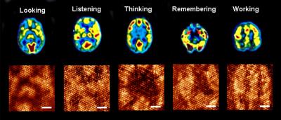
An international research team from Japan and Michigan Technological University have demonstrated a molecular circuit that can evolve continuously to solve complex problems that challenge today’s supercomputers. The massively parallel circuit contains a layer of molecular switches (monolayer) that simultaneously interact in a manner similar to the information processing performed by the neurons in the human brain. That is, they can evolve to tackle complex problems. Information processing circuits in digital computers, in contrast, are static, and operate serially. The abstract of a paper published Nature Physics, “Massively parallel computing on an organic molecular layer,” explains: “Modern computers operate at enormous speeds—capable of executing in excess of 10^13 instructions per second—but their sequential approach to processing, by which logical operations are performed one after another, has remained unchanged since the 1950s. In contrast, although individual neurons of the human brain fire at around just 10^3 times per second, the simultaneous collective action of millions of neurons enables them to complete certain tasks more efficiently than even the fastest supercomputer.”
Credit: Anirban Bandyopadhyay In the top half of the image above, you can see magnetic resonance images of the human brain during different functions. Below that, you can see similar evolving patterns have been generated on the molecular monolayer one after another. A snapshot of the evolving pattern for a particular brain function is captured using Scanning Tunneling Microscope at 0.68 V tip bias (scale bar is 6 nm). The molecular processor can also heal itself if there is a defect because of the self-organizing ability of the molecular monolayer. Like the brain, if a neuron dies, another neuron takes over its function. To prove that the parallel, dynamically reconfigurable processor works as claimed, the researchers have mimicked two natural phenomena in the molecular layer: heat diffusion and the evolution of cancer cells. The research was carried out by a team that includes Ranjit Pati, of the Michigan Technological University Department of Physics and Anirban Bandyopadhyay, National Institute for Materials Science, National Institute of Information and Communication Technology, Japan.
| ||
|
|
|
|
| |
|
Cranial Nerves
There are twelve cranial nerves altogether. These are twelve pairs of nerve tissue that arise from the brain itself, rather than the spinal cord. In order to innervate their target organs, they must exit/enter the cranium through openings in the skull (cranium). Hence, they are called cranial nerves. The nerves that arise from the spinal cord are called spinal nerves. What is the function of the cranial nerves? They play a very important role indeed. The first cranial nerve is the olfactory nerve. It is an entirely sensory nerve. There are specialised sensory receptive parts of the olfactory nerve which are located in the olfactory mucosa (a layer) of the upper parts of your nose. During breathing, air molecules attach to the olfactory mucosa and stimulate the olfactory receptors. Olfactory bulb cells then transmit electrical activity to other parts of the central nervous system via the olfactory tract. The smell that you are trying to identify is then interpreted by the brain. That’s why when you have a cold, you lose your sense of smell. The second cranial nerve is the optic nerve. The optic nerve starts from the cells of your retina inside your eye, where images that you see from the outside world are projected upside down. The cells then transmit the signals to your brain via the optic nerve. The optic nerve and tract has quite a complicated pathway. Anything that happens to this pathway (such as a tumour pressing the optic chiasm where the nerve fibres cross) will result in a certain part of an image (such as a vertical half of an image from one eye or the other, or even both) deleted from your vision. The brain will then interpret what you are seeing. The part of the brain that does this is the occipital lobe, the back portion. Does the 3rd cranial nerve have something to do with movements of the eye? Yes. Vision itself is transmitted through the 2nd cranial nerve, but the eye also has to move up and down and sideways. The 3rd cranial nerve is called the oculomotor nerve. This nerve is in charge of supplying the levator palpebrae superioris, which lifts your upper eyelid. It also supplies the superior rectus (moving your eye upward and inward, as in trying to look towards the inner contours of your eyebrow), medial rectus ( moving your eye inward towards the tip of your nose), inferior rectus (downward and inward) and inferior oblique (moving your eye upward and outward) muscles of the eye. The 3rd cranial nerve also has an autonomic component that constricts your pupils. A patient with a 3rd nerve palsy will then fail to raise his upper eyelid (ptosis), fail in all the movements of the eye described above and have a dilated pupil in the affected eye. The 4th cranial nerve (trochlear nerve) innervates the superior oblique muscle of the eye, which is responsible for turning the eye outwards and downward. The 6th cranial nerve (abducens nerve) is responsible for the lateral rectus muscle, which turns the eye outward. Naturally, a person with this palsy has a failure to look to his affected side. Description of the 5th and 7th cranial nerves? Are they large. Yes. The 5th (trigeminal) nerve is composed of three large branches – ophthalmic (V1), maxillary (V2) and mandibular (V3) branches. The ophthalmic branch is responsible for the sensation of the cornea and the area of skin from your forehead to your eyelids. The maxillary division gives sensation to the part of your face over the maxilla, nose, teeth of your upper jaw and palate. The mandibular branch takes care of your cheek, jaw, side of head and whether your first 2/3 of your tongue feels any pain/hot/cold. The mandibular branch is also responsible for the muscles of chewing. The 7th (facial nerve) is a very long nerve and partial to injury. It supplies the muscles for facial expression – furrowing your brow, closing your eye, smiling, puffing out your cheeks. It also supplies the taste component for the first 2/3rds of your tongue. If any part of this nerve is affected, you won’t be able to close your eye on the affected side, furrow your brow or smile. Which is the nerve responsible for hearing? The 8th (vestibulocochlear) nerve. True to its name, it has two main branches. The vestibular part is responsible for the position and movement of the head. The cochlear part is for hearing. Next we come to the nerves arising from the brainstem. The 9th (glossopharyngeal) nerve assists in swallowing. It also supplies the sensation and taste for the last 1/3 (back) part of your tongue. The 10th (vagus) nerve is the largest nerve in the body. It supplies your pharynx (in charge of constricting your throat), your larynx (voice box), the involuntary musculature of your bronchi inside your lungs, your heart, your intestinal tract, your liver and pancreas. The 11th (accessory) nerve supplies the muscles of your soft palate, pharynx and larynx and the great neck muscles. The 12th (hypoglossal) nerve supplies the muscles that move your tongue. | ||
|
|
|
|
| |
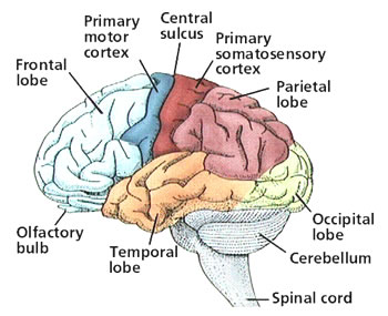
The cortex of each hemisphere has four lobes: the occipital, temporal, parietal, and frontal lobes. The occipital lobe controls vision. The temporal lobe controls sound and speech. The parietal lobe controls movement, touch, and recognition. And the frontal lobe controls thinking and planning 
| ||
|
|
|
|
| |
|
Tera-100 – Bull bullx super-node S6010/S6030(Bull SA)
System Name: Tera-100 System Family: Bull Bullx System Model: bullx super-node S6010/S6030 Computer: Bull bullx super-node S6010/S6030 Vendor: Bull SA Application Area: Defense Installation Year: 2010 Operating System: Linux Cores: 138368 Rmax(GFlops): 1050000 Rpeak(GFlops): 1254550 | ||
|
|
|
